About the contest
As part of the FEBS 60th anniversary celebrations this year, members of FEBS Constituent Societies and participants of the 48th FEBS Congress were invited to submit photographs taken with the use of bio-imaging tools (microscopes of any type) in the course of scientific work, for an exhibition showcasing “The Beauty Behind Biological Sciences”. The contest was organized by FEBS in collaboration with the Polish Biochemical Society and the Italian Society of Biochemistry and Molecular Biology.
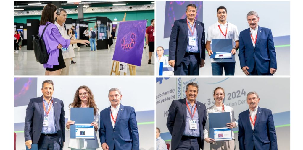
A large number of exceptional entries were received, and from these the judges had the hard task of selecting ten images that stood out for scientific significance, originality, and artistic impact. After selection, these ten images were printed at a large size for an exhibition in the centre of the poster area at the 48th FEBS Congress venue in Milano, from 29 June to 3 July 2024. All the event’s participants were able to enjoy the impact of these images as ‘art’ and were invited to vote for their favourite one (see top left picture). Voting was blind to submitter names and image descriptions.
Prizes
As a result of the participant voting, the following prizes were announced at the closing ceremony of the 48th FEBS Congress. Congratulations to all! Winners attending the closing ceremony were photographed receiving a prize certificate (see above).
First prize (€500): Román Martí Díaz, University of Murcia, Spain (pictured top right)
Second prize (€300): Jesús Soldán Hidalgo, University of Seville, Spain
Third prize (€200): Anna Richert, Nicolaus Copernicus University in Torun, Poland (pictured bottom left)
Distinction prizes (€100): Anna Pilatone, University of Padua, Italy (pictured bottom right) and Alessandra Lo Cicero, University of Palermo, Italy
We also congratulate the submitters of the five other outstanding images selected for the Congress exhibition: Alessandro Bevilacqua, Roberta Cascella, Karolina Krautforst, Wojciech Szlauer and Grzegorz Węgrzyn.
Images
The top five voted images are shown below, along with the image descriptions supplied with the entries:
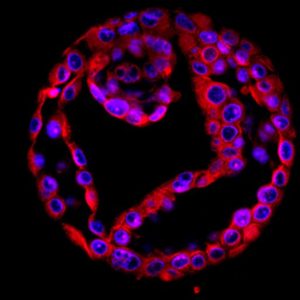
Yin & Yang in breast cancer mammospheres, Román Martí Díaz
Similar to the Yin & Yang principle, which symbolizes two opposing, complementary and interconnected forces, MCF7 breast cancer cells can switch from a differentiated to an invasive phenotype through an epithelial-mesenchymal transition (EMT). This cellular balance reflects a dynamic relationship in breast cancer. Confocal microscope Leica Stellaris used to image MCF7 breast cancer cells stained with 2 colors (E-cadherin in red and the nucleus in blue). Image brightness, size and resolution were adjusted using ImageJ software.
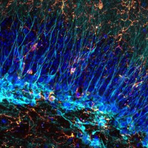
Atmospheric memory, Jesús Soldán Hidalgo
The image depicts the presence of microglial cells around doublecortin positive neurons in the dentate gyrus of a one-month-old C57BL/6 mouse. The nuclei of all cells are shown in blue, doublecortin positive neurons in cyan, microglia in orange. Some microglia positive for CD68 are illustrated in green. An immunofluorescence technique was developed on a 30 µm thickness brain section from a one-month-old C57BL/6 mouse. Antibodies against doublecortin, Iba1 and CD68, as well as DAPI, were used. The tissue was photographed under a LEICA Stellaris 8 – STED confocal microscope, with a 63X oil – immersion objective. A Z-stack was taken with a Z-step size of 0.80 µm and Maximum Intensity Projection was applied. Finally, image size was readjusted using Photoshop CS5.1 software.
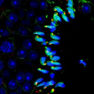
Parrot-painted spermatids, Anna Richert
The photo shows a cross-section of a mouse testicle in which we can observe the progressing process of spermatogenesis. Male generative cells are arranged in a cascade with increasing chromatin (blue) condensation, which is finally packed into a hook-shaped sperm head stabilized by actin (green) and myosin 6 (red). Testis fixed in 4% paraformaldehyde use to prepare cryo-tissue-sections. Permeabilization with 0.1% saponin and blocking with 1% BSA/PBS frozen sections. Incubation with the primary antibody (rabbit anti-Myosin 6, Proteintech, 1:200) in 0,15% BSA/PBS at 4 °C o/n and then with the secondary antibody (goat anti-rabbit IgG, Alexa Fluor Plus 594, Invitrogen, 1:100) in 0,1% BSA/PBS, RT for 2h. Actin filaments were visualized with Alexa Fluor 488 phalloidin (Invitrogen, 1:100). DNA was stained with Hoechst (Invitrogen, 1:5000). Images were acquired using confocal microscopy. Confocal microscope OLYMPUS FV 3000 (Software: OLYMPUS cellSens Dimension (Build 18987) RELEASE X64), Graphic software: light changes ImageJ, Fiji.
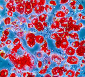
The fascinating world of adipocytes: a deep dive among droplets of energy, Anna Pilatone
Oil Red O staining of mature adipocytes containing small and large lipid droplets (LDs). These are highly dynamic organelles that resemble round bright red pearls. Initially surrounding nucleus as a crown, LDs then fuse each other until they fill the entire cytoplasm in one, almost, unique droplet of energy! Photos obtained using light microscope Leica ICC50W (20X magnification). PhotoPad Software Editor to modify brightness (+7), contrast (+5), saturation (+5), color (+5). No filter used. Oil Red O (ORO) is a fat soluble hydrophobic diazo dye that stains neutral fat, fatty acids and triglycerides, appearing red under light.
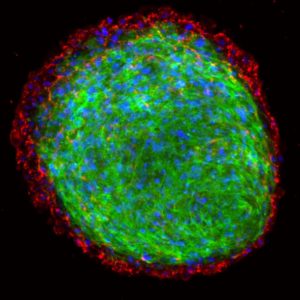
Endogenous extracellular matrix in primary tumor spheroid, Alessandra Lo Cicero
Tumorigenesis is a complex and dynamic process in which the extracellular matrix plays a leading role. Spheroids generated from primary tumor cells present an organized extracellular matrix. Collagen is mainly arranged around the 3D structure, mimicking simulating in vitro the in vivo tumor microenvironment conditions in solid tumors. Spheroids were stained with phalloidin-FITC (green), antibody anti-Collagen type I (red) and DAPI (blue) and analyzed with a confocal microscope (Olympus FV10i).